")
The term urolithiasis (or Kidney Stones or Urinary Tract Stones) refers to stone formation in the urinary tract. When it comes to congenital neural defects, urinary stones are associated with Spina Bifida as a secondary complication. Calculi in the urinary tract have long been recognized as a cause of morbidity in the patients with SB and other spinal cord defects. Although this complication is not common in the Western countries; however, it is associated with 5% of all congenital defects in the US [1]. The individuals may present with abdominal pain, dysuria, a burning sensation, haematuria and sometimes with vesicutaneus fistula (only few cases worldwide) [2].
How to diagnose urolithiasis, kidney stones or urinary tract stones in spina bifida?
After taking a complete history related to SB and associated symptoms of urolithiasis, the birth defect can be confirmed with the help of physical examination, lab investigations and radiology.
Physical examination
Findings on the physical examination of the patient with SB associated urinary stones vary from person to person. Most of the time, patients complain of pain in the urinary areas. If stones are suspected in the urethra, it can be palpated.
Laboratory Investigations
Urine analysis and urine culture are indicated for the patients with SB associated urolithiasis. Urine analysis is performed to measure the level of pH. A low pH on analysis is indicative of stone formation as the stones are formed at low pH levels. Urine culture is indicated to isolate the organism for the selection of antibiotics [3].
Imaging
Imaging is a useful tool for the confirmation of stones in the urinary tract and any other abnormality associated with urolithiasis in spina bifida. There are several tests in medical imaging to diagnose stones in urinary tract (urethra, bladder, ureters or in the kidneys).
Plain Radiographs
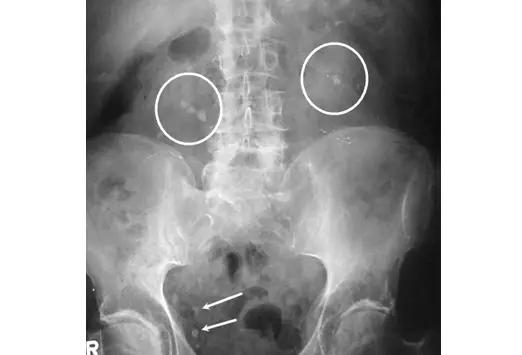
This procedure can diagnose more than 90% of the urinary stones. In cold areas, 60 to 70% of the stones are renal at the time of the diagnosis, but later they migrate to the urethra, bladder, ureters and to other urinary areas.
Ultrasonography
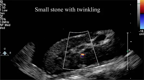
Ultrasonography is less useful in detecting the presence of ureteric stones, but when other advanced technologies are not available, it can be used. Ultrasonography has a significant role in the detection the congenital malformations found in SB and hydrocephalus.
Intravenous urography (I VU)
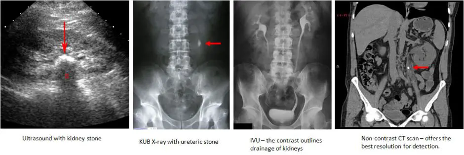
This technique is useful in detecting the mildly opaque stones or radiolucent stones (not picked up by plain radiography). Renal function tests of the patients with SB can be performed by this technique.
Treatment of Urolithiasis (Kidney Stones, Urinary Tract Stones) with Spina Bifida
Medical
The treatment plan for patients with SB should be medical or non-invasive because the complications of surgery and anaesthesia may worsen the illness (as patients with spina bifida have a neurological disorder and it is difficult to tolerate anaesthetic gases). A large quantity of fluid intake should be advised to ensure satisfactory urinary output, especially for those individuals living in hot climates. This high fluid intake is required for life to minimize the chances of reoccurrence.
A major complaint of the patients of spina bifida associated urolithiasis is pain. This can be treated with narcotics and antispasmodic drugs. Antibiotic therapy must be advised to avoid possible urinary tract infection (UTI). Sometimes bacteria trapped in the calculi are not easily accessible so lengthy antibiotic therapy should be advised. It is advisable to reduce the use of calcium rich products such as milk and cheese to reduce the possible chances of calcified stones. In the same manner, also reduce the use of oxalate rich products such as nuts and cola drinks [4]. Potassium citrate is a calcium stone inhibitor and it may be helpful to reduce the development of urinary tract stones or kidney stones.
Doctors at Children’s Hospital of PIttsburgh of UPMC have come up a unique and cutting-edge solution for Arianna Evans, a 16-year-old spina bifida patient in renal failure who frequently needs dialysis.
Lithotripsy (Extra corporeal shock wave lithotripsy)
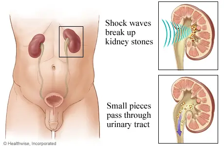
This procedure is a non-invasive and safe mode of treatment for the patients with spina bifida associated urinary stones. At the start, it was only performed on the adult patients, but with the passage of time it has been used on children successfully [5]. The stone is localised with an ultrasound scan and shock waves are applied at the site of stones. Lithotripsy is recommended after ensuring that there is no obstruction in the urinary tract that may block the passage of the broken stones. The stone should be single as multiple stones and obesity are contraindications of lithotripsy.
Surgery
It is not considered safe to treat the SB patients with a surgical procedure; however, in some conditions it is necessary to remove stone surgically (as in the case of patients with urinary obstruction). Several surgical procedures are available for the removal of SB associated urinary stones. Some of these procedures are:
- Minimal invasive surgery
- Open surgery [6]
Prevention and Follow-up
- It is necessary for patients of spina bifida to visit their consultant regularly according to the scheduled follow up.
- Regular ultrasounds or other means of diagnosis may be indicated.
- Careful dietary intake can also help the spina bifida patient stop the formation of urinary stones.
References
- Schwartz BF, Stoller ML. The vesical calculus. Urol Clin North Am 2000; 27:333.
- Hanif GM, Sameer PJ, Kirtipal NV, Hemang B. Giant Vesical calculus presenting as a vesicocutaneous fistula. Urol Int 48:115.
- Davenport M. ABC of general surgery in hildren: acute abdominal pain n children. BMJ 1996;312:498-501.
- Pak CYC, Fuller C, Sakhaee K, Preminger GM, Britton F. Long term treatment of calcium nephrolithiasis with potassium citrate. J Urol 1985; 134:11-19.
- Kroovand RL, Harrison LH, McCullough DL. Extracorporeal shock wave lithotripsy in childhood. J Urol 1987; 138:1106-8.
- Mshelbwala PM, Ameh EA, and Mbibu HN. Urinary stones in children: Review article. Niger J Surg Res 2005; 79(3–4):238-43.
Related Articles:
- 6 Complications of Bladder Augmentation in Spina Bifida
- 5 Facts about Neurogenic Bladder Treatment
- 15 Health Care Issues for Adults living with Spina Bifida
- 6 Disorders Associated with Spina Bifida
- Lordosis: Causes, Symptoms, Diagnosis, Treatment & Exercises
- Occupational & Physical Therapy Treatment for Spina Bifida
We will add your organization!